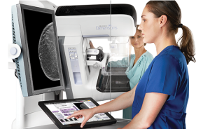
 Patient wellness and continuum of care are Assured Imaging’s main focus. Both our mobile coaches and our fixed locations provide the latest in cutting-edge, proven technology.
• Assured Imaging offers both mobile digital 3D and 2D mammogram screenings.
• 1 in 8 women will be diagnosed with breast cancer.
• Women 40 and older should get a mammogram screening once every year.
• Nearly all breast cancer is successfully treated if detected early!
3D Tomosynthesis Breast Imaging
3D mammography enables physicians to see masses and distortions more clearly. Tomosynthesis obtains multiple images from a variety of angles through the entire breast. Even fine details become obvious because they’re no longer hidden by surrounding tissue as they would be with a two-dimensional mammogram.
- 3D mammography allows doctors to examine your breast tissue layer by layer. So, instead of viewing all of the complexities of your breast tissue in a flat image, as with traditional 2D mammography, fine details are more visible and no longer hidden by the tissue above or below.
- 3D mammography detects 41% more invasive breast cancers and reduces the chance that you will need to be called back for additional views by up to 40%.
Prepare for Your Visit
How To Prepare
- Talk to your health care professional about any concerns you have regarding the need for the test you have scheduled, it’s risks, how it will be performed, or what the results will mean to you.
- The day of your mammogram wear a two-piece outfit so that it is easy to undress above the waist.
- Please do not wear lotion, powder, perfume or deodorant to your appointment. Wearing these items can make mammograms more difficult to read and may corrupt equipment. If you must wear deodorant, moist towelettes will be available so you can remove it before your exam.
When breast cancer is detected early treatment is easier and more successful. For that reason, some experts recommend that women over 20 perform a monthly breast self exam to look for new lumps and other changes. The self exam does have limitations, however,and is not a substitute for regular breast examinations from your doctor and screening mammograms. If you do perform monthly exams, it’s best to do them 3-5 days after your period ends, when your breasts are the least tender and lumpy.
How to perform a self breast exam: First, lie on your back. Place your right hand behind your head. With the middle fingers of your left hand, gently yet firmly press down using small motions to examine the entire right breast. Then, while sitting or standing, examine your armpit, an important area that is commonly overlooked. Breast tissue extends to this area, so it is important that it be included in every breast exam. Gently squeeze the nipple, checking for discharge. Repeat the process on the left breast.
Although some women find it easiest to do the exam in the shower when the skin is soft and wet, you are more likely to examine all of the breast tissue if you are lying down. Next, stand in front of a mirror with your arms by your side. Look at your breasts directly, as well as in the mirror. Search for changes in skin texture, such as dimpling, puckering, indentations, and skin with an “orange peel-like texture”. Look for changes in shape, contour, and inversion of the nipples. Finally, perform the exam again, this time with your arms raised above your head.
Immediately discuss any changes you find with your doctor. Most women have some naturally occurring lumps in their breasts, but it’s important that you become familiar with the way your breasts normally feel, so you can be aware of any new changes. Though the American Cancer Society considers self examinations to be optional, it’s a good idea to talk to your health care provider about what is right for you.
References:
US Preventive Services Task Force, Screening for Breast Cancer Recommendations and Rationale
Guide to Clinical Preventive Services: Third Edition (2000-2003).
Rockville, Maryland US Department of Health and Human Services, Public Health Service, Agency for Health Care Policy and Research; 2002.
What is your risk of breast cancer? Do antiperspirants increase the risk of breast cancer? Can breast cancer be prevented? When it comes to breast health, what you don’t know can hurt you. Misinformation can keep you from recognizing and minimizing your own risk of breast cancer and receiving the best possible care. Arm yourself with the facts.
Most Common Breast Cancer Myths
While it’s true that the risk of breast cancer increases as you grow older, breast cancer can occur at any age. From birth to age 39, one woman in 231 will get breast cancer (<0.5% risk); from age 40-59, the risk is one in 25 (4% risk) and from age 60-79, the risk is one in 15 (nearly 7%). If a woman were to live to age 90, her risk of getting breast cancer over the course of her entire lifetime is one in 7, with an overall lifetime risk of 14.3%.
Getting breast cancer is never a certainty even if you have one of the stronger risk factors, like a breast cancer gene abnormality. Of women with a BRCA1 or BRCA2 inherited genetic abnormality, 40-80% will develop breast cancer over their lifetime and 20-60% won’t. All other risk factors are associated with a much lower probability of being diagnosed with breast cancer.
Every woman has some risk of breast cancer, in fact increasing age is the biggest single risk factor for getting the disease. About 80% of women who get breast cancer have no known family history of the disease. If you do have a family history of breast cancer and are concerned, discuss it with your physician or a genetic counselor. You may be worrying needlessly.
A history of breast cancer in both your mother’s and your father’s family will influence your risk equally. That’s because half of your genes come from your mother, half from your father. However, a man with a breast cancer gene abnormality is less likely to develop breast cancer than a woman with a similar gene. So, if you want to learn more about your father’s family history, you have to look mainly at the women on your father’s side.
There is no evidence that the active ingredient in antiperspirants influences breast cancer risk. The supposed link between breast cancer and antiperspirants is based on misinformation about anatomy and a misunderstanding of breast cancer.
Modern day birth control pills contain a low dose of the hormones estrogen and progesterone. Many research studies show no association between birth control pills and an increased risk of breast cancer. However, one study that combined the results of many different studies did show an association between birth control pills and a very small increase in risk. The study also showed that this slight increase in risk decreased over time. So after 10 years, birth control pills were not associated with an increase in risk. Birth control pills also have benefits, such as decreasing ovarian and endometrial cancer risk; relieving menstrual disorders, pelvic inflammatory disease, and ovarian cysts; and improving bone mineral density As with any medicine, you have to weigh the risks and benefits and decide what is best for you.
Thus far, studies have not been able to demonstrate a clear connection between eating high-fat foods and a higher risk of breast cancer, though the issue is still being investigated. Regardless, avoidance of high-fat foods is a healthy choice for a multitude of reasons: to lower “bad” cholesterol (low-density lipoproteins), increase “good” cholesterol (high-density lipoproteins), to make more room in your diet for healthier foods, and to help you control your weight. And excess body weight is a risk factor for breast cancer because the extra fat increases the production of estrogen outside the ovaries and adds to the overall level of estrogen in the body. If you are already overweight, or have a tendency to gain weight easily, avoiding high-fat foods is strongly advised.
Digital mammography or high quality film-screen mammography is the most reliable way to find breast cancer as early as possible, when it is most curable. By the time a breast cancer can be felt, it is usually larger than the average size of a cancer first found on mammography. However, breast examinations by you and your health care provider are still very important. About 25% of breast cancers are found only on breast examination (not on the mammogram), about 35% are found on mammography alone, and 40% are found by both physical exam and mammography. It makes good breast health sense to keep all your bases covered.
There are several effective ways to reduce the risk of breast cancer in women with high risk factors. Options include lifestyle changes, such as minimizing alcohol consumption, stopping smoking, and exercising regularly. Another option is medication (Tamoxifen, also called Nolvadex) and in cases of very high risk, prophylactic mastectomy surgery or prophylactic ovary removal may be offered. However, be sure that you consult with a physician or genetic counselor before you make assumptions about your level of risk. If you have any questions about this content, please let me know.
Every woman has some risk of breast cancer, in fact increasing age is the biggest single risk factor for getting the disease. About 80% of women who get breast cancer have no known family history of the disease. If you do have a family history of breast cancer and are concerned, discuss it with your physician or a genetic counselor. You may be worrying needlessly.
Patient FAQ
A mammogram is a low-dose x-ray exam of the breasts to look for changes that are not normal. The results are recorded on x-ray film or directly into a computer for a doctor called a radiologist to examine.
A mammogram allows the doctor to have a closer look for changes in breast tissue that cannot be felt during a breast exam. It is used for women who have no breast complaints and for women who have breast symptoms, such as a change in the shape or size of a breast, a lump, nipple discharge, or pain. Breast changes occur in almost all women. In fact, most of these changes are not cancer and are called “benign,” but only a doctor can know for sure. Breast changes can also happen monthly, due to your menstrual period.
A high-quality mammogram plus a clinical breast exam, an exam done by your doctor, is the most effective way to detect breast cancer early. Finding breast cancer early greatly improves a woman’s chances for successful treatment.
Like any test, mammograms have both benefits and limitations. For example, some cancers can’t be found by a mammogram, but they may be found in a clinical breast exam.
Checking your own breasts for lumps or other changes is called a breast self-exam (BSE). Studies so far have not shown that BSE alone helps reduce the number of deaths from breast cancer. BSE should not take the place of routine clinical breast exams and mammograms.
If you choose to do BSE, remember that breast changes can occur because of pregnancy, aging, menopause, menstrual cycles, or from taking birth control pills or other hormones. It is normal for breasts to feel a little lumpy and uneven. Also, it is common for breasts to be swollen and tender right before or during a menstrual period. If you notice any unusual changes in your breasts, contact your doctor.
You stand in front of a special x-ray machine. The person who takes the x-rays, called a radiologic technician, places your breasts, one at a time, between an x-ray plate and a plastic plate. These plates are attached to the x-ray machine and compress the breasts to flatten them. This spreads the breast tissue out to obtain a clearer picture. You will feel pressure on your breast for a few seconds. It may cause you some discomfort; you might feel squeezed or pinched. This feeling only lasts for a few seconds, and the flatter your breast, the better the picture. Most often, two pictures are taken of each breast - one from the side and one from above. A screening mammogram takes about 20 minutes from start to finish.
- Screening mammogramsare done for women who have no symptoms of breast cancer. It usually involves two x-rays of each breast. Screening mammograms can detect lumps or tumors that cannot be felt. They can also find microcalcifications (my-kro-kal-si-fi-KAY-shuns) or tiny deposits of calcium in the breast, which sometimes mean that breast cancer is present.
- Diagnostic mammogramsare used to check for breast cancer after a lump or other symptom or sign of breast cancer has been found. Signs of breast cancer may include pain, thickened skin on the breast, nipple discharge, or a change in breast size or shape. This type of mammogram also can be used to find out more about breast changes found on a screening mammogram, or to view breast tissue that is hard to see on a screening mammogram. A diagnostic mammogram takes longer than a screening mammogram because it involves more x-rays in order to obtain views of the breast from several angles. The technician can magnify a problem area to make a more detailed picture, which helps the doctor make a correct diagnosis.
A digital mammogram also uses x-rays to produce an image of the breast, but instead of storing the image directly on film, the image is stored directly on a computer. This allows the recorded image to be magnified for the doctor to take a closer look. Digital mammography may offer these benefits:
- Long-distance consultations with other doctors may be easier because the images can be shared by computer.
- Slight differences between normal and abnormal tissues may be more easily noted.
- The number of follow-up tests needed may be fewer.
- Fewer repeat images may be needed, reducing exposure to radiation.
The National Cancer Institute recommends:
- Women 40 years and older should get a mammogram every 1 to 2 years.
Women who have had breast cancer or other breast problems or who have a family history of breast cancer might need to start getting mammograms before age 40, or they might need to get them more often. Talk to your doctor about when to start and how often you should have a mammogram.
The radiologist will look at your x-rays for breast changes that do not look normal and for differences in each breast. He or she will compare your past mammograms with your most recent one to check for changes. The doctor will also look for lumps and calcifications.
- Lump or mass.The size, shape, and edges of a lump sometimes can give doctors information about whether or not it may be cancer. On a mammogram, a growth that is benign often looks smooth and round with a clear, defined edge. Breast cancer often has a jagged outline and an irregular shape.
- A calcification is a deposit of the mineral calcium in the breast tissue. Calcifications appear as small white spots on a mammogram. There are two types:
- Macrocalcificationsare large calcium deposits often caused by aging. These usually are not a sign of cancer.
- Microcalcificationsare tiny specks of calcium that may be found in an area of rapidly dividing cells.
If calcifications are grouped together in a certain way, it may be a sign of cancer. Depending on how many calcium specks you have, how big they are, and what they look like, your doctor may suggest that you have other tests. Calcium in the diet does not create calcium deposits, or calcifications, in the breast.
If you have a screening test result that suggests cancer, your doctor must find out whether it is due to cancer or to some other cause. Your doctor may ask about your personal and family medical history. You may have a physical exam. Your doctor also may order some of these tests:
- Diagnostic mammogram, to focus on a specific area of the breast
- Ultrasound, an imaging test that uses sound waves to create a picture of your breast. The pictures may show whether a lump is solid or filled with fluid. A cyst is a fluid-filled sac. Cysts are not cancer. But a solid mass may be cancer. After the test, your doctor can store the pictures on video or print them out. This exam may be used along with a mammogram.
- Magnetic resonance imaging (MRI), which uses a powerful magnet linked to a computer. MRI makes detailed pictures of breast tissue. Your doctor can view these pictures on a monitor or print them on film. MRI may be used along with a mammogram.
- Biopsy, a test in which fluid or tissue is removed from your breast to help find out if there is cancer. Your doctor may refer you to a surgeon or to a doctor who is an expert in breast disease for a biopsy.
Women with breast implants should also have mammograms. A woman who had an implant after breast cancer surgery in which the entire breast was removed (mastectomy) should ask her doctor whether she needs a mammogram of the reconstructed breast.
If you have breast implants, be sure to tell your mammography facility that you have them when you make your appointment. The technician and radiologist must be experienced in x-raying patients with breast implants. Implants can hide some breast tissue, making it harder for the radiologist to see a problem when looking at your mammogram. To see as much breast tissue as possible, the x-ray technician will gently lift the breast tissue slightly away from the implant and take extra pictures of the breasts.
First, check with the place you are having the mammogram for any special instructions you may need to follow before you go. Here are some general guidelines to follow:
- If you are still having menstrual periods, try to avoid making your mammogram appointment during the week before your period. Your breasts will be less tender and swollen. The mammogram will hurt less and the picture will be better.
- If you have breast implants, be sure to tell your mammography facility that you have them when you make your appointment.
- Wear a shirt with shorts, pants, or a skirt. This way, you can undress from the waist up and leave your shorts, pants, or skirt on when you get your mammogram.
- Don’t wear any deodorant, perfume, lotion, or powder under your arms or on your breasts on the day of your mammogram appointment. These things can make shadows show up on your mammogram.
- If you have had mammograms at another facility, have those x-ray films sent to the new facility so that they can be compared to the new films.
Although they are not perfect, mammograms are the best method to find breast changes that cannot be felt. If your mammogram shows a breast change, sometimes other tests are needed to better understand it. Even if the doctor sees something on the mammogram, it does not mean it is cancer.
As with any medical test, mammograms have limits. These limits include:
- They are only part of a complete breast exam.Your doctor also should do a clinical breast exam. If your mammogram finds something abnormal, your doctor will order other tests.
- Finding cancer does not always mean saving lives.Even though mammography can detect tumors that cannot be felt, finding a small tumor does not always mean that a woman’s life will be saved. Mammography may not help a woman with a fast growing cancer that has already spread to other parts of her body before being found.
- False negatives can happen.This means everything may look normal, but cancer is actually present. False negatives don’t happen often. Younger women are more likely to have a false negative mammogram than are older women. The dense breasts of younger women make breast cancers harder to find in mammograms.
- False positives can happen.This is when the mammogram results look like cancer is present, even though it is not. False positives are more common in younger women, women who have had breast biopsies, women with a family history of breast cancer, and women who are taking estrogen, such as menopausal hormone therapy.
- Mammograms (as well as dental x-rays and other routine x-rays) use very small doses of radiation.The risk of any harm is very slight, but repeated x-rays could cause cancer. The benefits nearly always outweigh the risk. Talk to your doctor about the need for each x-ray. Ask about shielding to protect parts of the body that are not in the picture. You should always let your doctor and the technician know if there is any chance that you are pregnant.
7717 N Hartman Ln
Tucson, AZ 85743
- (520) 744-6121
- (888) 233-6121
- (520) 572-7138
- [email protected]
Assured Imaging Women’s Wellness is the leading provider of mobile digital mammography in the United States. With a footprint that covers more than 60 locations across 16 states we are doing more to promote early detection of breast cancer than any other similar provider in the country.
Copyright © 2019 Assured Imaging
Assured Imaging is a subsidiary of United Medical Imaging. Who is UMI?


